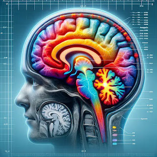
Post-Traumatic Stress Disorder (PTSD) is a debilitating condition that affects millions of people worldwide. While traditionally associated with war veterans, PTSD can arise from any traumatic event, including accidents, natural disasters, and personal assaults. Despite its prevalence, PTSD has long been misunderstood and challenging to treat effectively. However, advances in neuroimaging are revolutionizing our understanding of PTSD, providing new insights into its neurobiological underpinnings and paving the way for more effective treatments.
Understanding PTSD: The Role of Neuroimaging
Neuroimaging refers to various techniques used to visualize the structure and function of the brain. Functional magnetic resonance imaging (fMRI) is one of the most widely used neuroimaging tools in PTSD research. fMRI measures brain activity by detecting changes in blood flow, allowing researchers to see which areas of the brain are more active during specific tasks or in response to certain stimuli.
Recent studies using fMRI have identified significant differences in the brain activity of individuals with PTSD compared to those without the disorder. These differences are particularly pronounced in the amygdala, prefrontal cortex, and hippocampus – areas of the brain involved in emotion regulation, fear processing, and memory. Understanding these abnormalities is crucial for developing targeted therapies that address these specific brain changes.
The Amygdala: The Brain’s Fear Center
The amygdala is a small, almond-shaped structure located deep within the brain. It plays a critical role in processing emotions, particularly fear. In individuals with PTSD, the amygdala is often hyperactive, leading to exaggerated fear responses and heightened anxiety.
A study published in Nature Neuroscience in 2023 highlighted this hyperactivity in the amygdala of PTSD patients. Researchers found that when exposed to trauma-related stimuli, individuals with PTSD showed significantly greater amygdala activation compared to healthy controls. This heightened activity was correlated with the severity of their PTSD symptoms, suggesting that amygdala hyperactivity is a key feature of the disorder.
Understanding the role of the amygdala in PTSD has important implications for treatment. Therapies that target amygdala hyperactivity, such as exposure therapy and certain medications, may help reduce fear responses and alleviate symptoms. Additionally, neurofeedback techniques, which train individuals to regulate their own brain activity, have shown promise in normalizing amygdala function and reducing PTSD symptoms.
The Prefrontal Cortex: Regulating Emotions and Behavior
The prefrontal cortex (PFC) is involved in higher-order cognitive functions such as decision-making, impulse control, and emotion regulation. In individuals with PTSD, the PFC often shows reduced activity, impairing their ability to regulate emotions and control fear responses.
A 2022 study published in The Journal of Neuroscience found that individuals with PTSD exhibited decreased activity in the ventromedial prefrontal cortex (vmPFC) – a region of the PFC associated with fear extinction and emotion regulation. This reduced activity was linked to difficulties in suppressing fear responses and regulating negative emotions.
These findings suggest that therapies aimed at enhancing PFC function could be beneficial for PTSD patients. Cognitive-behavioral therapy (CBT), which focuses on changing negative thought patterns and behaviors, has been shown to increase PFC activity and improve emotion regulation. Additionally, transcranial magnetic stimulation (TMS), a non-invasive procedure that uses magnetic fields to stimulate nerve cells, has been found to enhance PFC function and reduce PTSD symptoms.
The Hippocampus: Memory and Contextual Processing
The hippocampus is a crucial brain structure involved in memory formation and contextual processing. In PTSD, the hippocampus often shows reduced volume and impaired function, contributing to difficulties in distinguishing between safe and threatening environments and leading to persistent, intrusive memories of the traumatic event.
A groundbreaking study published in Biological Psychiatry in 2021 used high-resolution fMRI to examine hippocampal function in PTSD patients. The researchers found that individuals with PTSD had significantly reduced hippocampal volume and impaired connectivity between the hippocampus and other brain regions involved in memory and emotion regulation. These abnormalities were associated with the severity of their PTSD symptoms, highlighting the critical role of the hippocampus in the disorder.
Understanding hippocampal dysfunction in PTSD has important implications for treatment. Interventions that enhance hippocampal function, such as mindfulness-based stress reduction (MBSR) and physical exercise, have been shown to improve PTSD symptoms. Additionally, pharmacological treatments that promote neurogenesis (the growth of new neurons) in the hippocampus, such as selective serotonin reuptake inhibitors (SSRIs), have been found to be effective in reducing PTSD symptoms.
Emerging Neuroimaging Techniques
While fMRI has been instrumental in advancing our understanding of PTSD, other neuroimaging techniques are also providing valuable insights. Diffusion tensor imaging (DTI), for example, is a type of MRI that measures the integrity of white matter tracts in the brain. White matter tracts are the pathways that connect different brain regions and facilitate communication between them.
A 2022 study published in NeuroImage: Clinical used DTI to examine white matter integrity in PTSD patients. The researchers found that individuals with PTSD had reduced integrity in several key white matter tracts, including those connecting the amygdala, PFC, and hippocampus. These findings suggest that PTSD is associated with disruptions in brain connectivity, which may contribute to the symptoms of the disorder.
Positron emission tomography (PET) is another neuroimaging technique that has been used to study PTSD. PET scans measure the distribution of specific neurotransmitters and receptors in the brain, providing insights into the neurochemical changes associated with PTSD. A recent PET study published in Molecular Psychiatry found that individuals with PTSD had altered levels of certain neurotransmitters, such as serotonin and dopamine, which are involved in mood regulation and stress response.
The Future of PTSD Treatment: Personalized and Precision Medicine
The insights gained from neuroimaging studies are paving the way for more personalized and precision-based approaches to PTSD treatment. By identifying specific brain abnormalities and neurochemical changes associated with PTSD, researchers can develop targeted therapies that address these underlying issues.
For example, neuroimaging can help identify which patients are likely to respond to certain treatments. A study published in JAMA Psychiatry in 2023 used fMRI to predict which PTSD patients would respond to exposure therapy. The researchers found that patients with greater pre-treatment activity in the vmPFC were more likely to benefit from the therapy, suggesting that fMRI could be used to tailor treatment plans to individual patients.
Additionally, neuroimaging can help monitor treatment progress and outcomes. By regularly imaging the brain during treatment, clinicians can assess whether the therapy is effectively targeting the relevant brain regions and making the desired changes in brain activity. This approach allows for adjustments to be made in real-time, ensuring that patients receive the most effective treatment possible.
Challenges and Ethical Considerations
While the advances in neuroimaging are exciting, there are also challenges and ethical considerations to keep in mind. Neuroimaging studies are expensive and resource-intensive, which can limit their accessibility and scalability. Additionally, interpreting neuroimaging data requires specialized expertise, and there is still much we do not understand about the brain’s complex functioning.
Ethically, it is important to consider the implications of using neuroimaging to diagnose and treat PTSD. There are concerns about privacy and the potential for misuse of neuroimaging data. It is crucial to establish guidelines and safeguards to protect patients’ rights and ensure that neuroimaging is used responsibly and ethically.
Advances in neuroimaging are transforming our understanding of PTSD, providing unprecedented insights into the neurobiological changes associated with the disorder. By identifying specific brain abnormalities and neurochemical changes, researchers are developing more targeted and effective treatments for PTSD. While there are challenges and ethical considerations to address, the future of PTSD treatment looks promising, with the potential for personalized and precision-based approaches that offer hope and healing to those affected by this debilitating condition.
As we continue to explore the brain’s intricate workings, we move closer to
unlocking the mysteries of PTSD and developing interventions that can truly make a difference in the lives of those who suffer from it. The journey is ongoing, but with each discovery, we gain a deeper understanding and a renewed commitment to alleviating the burden of PTSD.
Reference List
Haris, E. M., Bryant, R. A., Williamson, T., & Korgaonkar, M. S. (2023). Functional connectivity of amygdala subnuclei in PTSD: a narrative review. Molecular Psychiatry, 28(9), 3581–3594. https://doi.org/10.1038/s41380-023-02291-w
Winter, N. R., Blanke, J., Leenings, R., Ernsting, J., Fisch, L., Sarink, K., Barkhau, C., Emden, D., Thiel, K., Flinkenflügel, K., Winter, A., Goltermann, J., Meinert, S., Dohm, K., Repple, J., Gruber, M., Leehr, E. J., Opel, N., Grotegerd, D., . . . Hahn, T. (2024). A Systematic evaluation of Machine Learning–Based biomarkers for major depressive Disorder. JAMA Psychiatry. https://doi.org/10.1001/jamapsychiatry.2023.5083
Li, L., Pan, N., Zhang, L., Lui, S., Huang, X., Xu, X., Wang, S., Lei, D., Li, L., Kemp, G. J., & Gong, Q. (2020). Hippocampal subfield alterations in pediatric patients with post-traumatic stress disorder. Social Cognitive and Affective Neuroscience, 16(3), 334–344. https://doi.org/10.1093/scan/nsaa162
.webp)

0 comments